Localización y tamaño del riñón en perros en radiografías abdominales
Para cada gato, los datos recopilados incluyeron la señalización y la historia, incluida la duración del estreñimiento/obstinación (tenesmo sin defecación) antes de la presentación. El diagnóstico de megacolon se realizó en un inicio con base en la historia clínica (primordialmente estreñimiento prolongado), los hallazgos clínicos correspondientes y el examen clínico (p. ej., palpación abdominal). Proyección dorsoventral del tórax de un perro sano.Los pliegues de piel crean bandas de densidad tejido blando queaparecen a los dos lados del tórax (flechas). En el hemitórax derechoel contraste de grises entre la zona del costado y la zona medial al plieguees tan marcado que podría confundirse con un neumotórax. Uncriterio de diferenciación es que el aire contenido en elespacio pleural continúa estático, al tiempo que elgas contenido en los pulmones se desplaza con la inspiración y la espiración.
 After performing X-rays, the staff discovered she had a number of urinary stones that needed to be eliminated. Our Board-certified Veterinary Surgeon, Dr. Lynne Snow, carried out a cystotomy to remove the stones from Scarlett’s bladder. She has since recovered and is again to her regular self together with her loving household. 24-hour hospital with an emergency vet in Toms River and board-certified docs prepared to help.
Vet-Ray’s AI is artificial intelligence built on thousands of companion animal cases.
Like a human hospital, sufferers with important, life-threatening injuries or conditions are handled with the highest precedence. After the pictures are taken, our radiologists will evaluate them and consult along with your pet’s care group. A physician will meet with you to debate your pet’s prognosis and therapy plan. Our staff additionally companions with your loved ones veterinarian to provide one of the best therapy options on your pet. Like different veterinary companies, X-rays are sometimes more expensive in urban and extremely populated places than in rural locations. And the value of X-rays can range by the veterinary practice in the same metropolis, so you may profit from shopping round.
Pet Insurance Providers with X-Ray Coverage
It’s usually essential in your vet to order X-rays for suspected orthopedic issues, such as X-rays of dogs with hip dysplasia. This X-ray provides your vet a view into your dog’s hip to see how this hereditary condition has progressed and can help your vet decide the best therapy course. Karu’s Animal Centre & Surgery is a humble vet clinic located in Pudu, Cheras. The medical doctors are described as pleasant, helpful and experienced by their shoppers. Most importantly, they're available for late-night emergencies, so name the doctor first when you require immediate assistance. Founded in yr 2011 by Dr. Thiba Rajoo, the group from Ministre’ Of Pets Veterinary Clinic is pleasant, attentive and all the time goes the extra mile to assist the animals, even the rescued ones!
Cyberlynx Animal Clinic
Kota Damansara Veterinary Centre is among the branches under Veterinary Essential Services. The clinic opens till 10pm on weekdays, which is perfect for many pet parents who wish to visit the vet after work. Our furry pals can’t inform us when they're experiencing discomfort. But they might show indicators that can help us see that they don’t feel nicely. If you are feeling your pet is experiencing any of those signs, contact your beloved ones veterinarian or come directly to MedVet Pittsburgh if it’s an emergency. Best Friends Pet Clinic - free service to you in connecting with local vets. All service suppliers are independent and we do not warrant or guarantee any service carried out or offered.
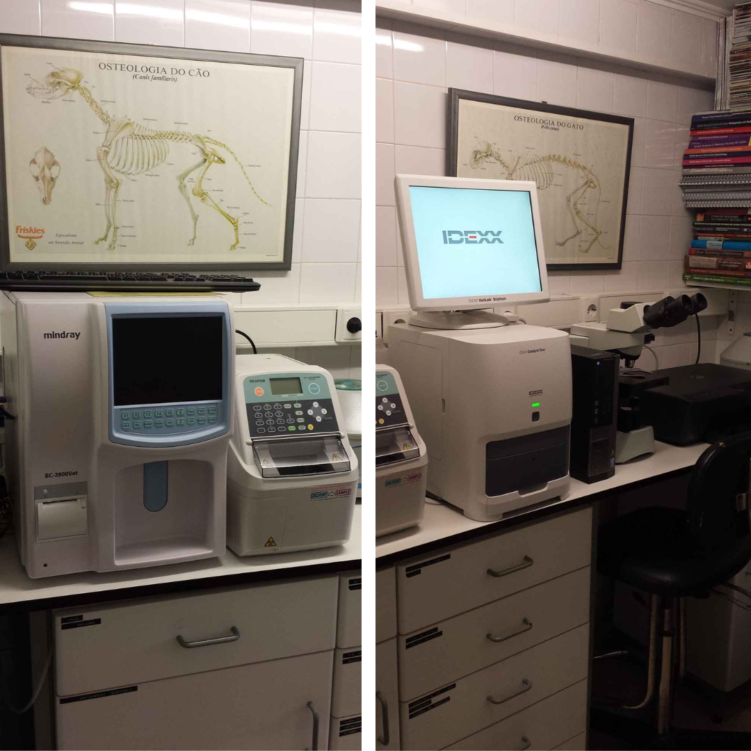
Similarly, lesions affecting the pylorus may be more evident on a left lateral radiographic examination of the stomach than on a right lateral. For this cause, a set of three views of the abdomen is now normal in most American veterinary educating hospitals. DR systems have been developed that do not require a cable to speak between the detector and the pc processing the info into an image. The cable has been changed by wireless communication on specified electrical magnetic frequencies which may be unlikely to be interfered with by other electromagnetic gadgets such as cell telephones and electronic gear.
Magnetic Resonance Imaging
Radiographs are made using a specialized kind of vacuum tube that produces x-rays. Kilovoltage potential (kVp) is the highest potential voltage achieved at any given kV setting. Although ultrasound can be utilized to gauge most delicate tissues, the guts and belly organs are essentially the most frequently scanned in veterinary clinics. The construction and performance of the center and its valves could be evaluated by this procedure. There are limitations to ultrasonography, because it can't be used to scan gas-filled (lungs, intestine) or bony tissues. The VMTH is equipped with state-of-the-art radiography methods that seize high quality photographs.
This could be given intravenously to examine organs like the kidneys or coronary heart, or by mouth to look at the digestive tract. A series of x-rays is taken after the dye is given, which will define the organs where the dye collects. Digital recording is in widespread use, however radiographic photographs have historically been, and a few still are, saved on specifically optimized movie. However, even one of the best silver halide film is comparatively insensitive to x-rays.
Complications of X-Rays for Dogs
If you’re considering giving CBD to your canine, you need to start by talking to your veterinarian to guarantee that your canine is an effective candidate for CBD. The results of CBD are noticeable at variable instances, relying on the product you employ, the route of administration, the dose required for your dog’s ailment, and the ailment itself. Before purchasing a vet x-ray machine in your clinic, you must take all of the above issues and communicate with a educated vendor. An experienced vet x-ray machine vendor will have the power to consider your whole current needs and future progress plans and counsel the finest possible machine in your explicit circumstances. It can be useful to document the settings used for every publicity, both on the system or by hand, so with time, we are in a position to begin to understand our machine and what settings work well for sure photographs. When serious about radiation security, both the affected person and the operator, at all times use the lowest potential settings wanted to gain the diagnostic image.
Are X-rays for dogs worth it?
However, these procedures often require common anesthesia or sedation and are more labor-intensive and dear. Be positive to talk together with your veterinarian in regards to the dangers and benefits of contrast radiography compared to alternate options such as ultrasound, MRI, and CT. Some veterinary services may base their costs on the scale of the canine or the location of the X-ray (e.g., dental vs. abdomen), while others may have a fixed fee regardless of the view. Even proficient individuals can miss lesions that are unfamiliar to them, or so-called "lesions of omission." A lesion of omission is one during which a structure or AnáLises VeterináRias organ usually depicted on the image is lacking. A good instance of that is the absence of 1 kidney or the spleen on an stomach radiograph. Therefore, specific consideration to systematic analysis of the image is essential. It is maybe best to begin interpretation of the image in an space that's not of primary concern.